Monday, August 30, 2010


Advanced Cell Technology, Inc., a biotechnology company applying cellular technology in the field of regenerative medicine, announced today that the U.S. Food and Drug Administration (FDA) has granted orphan drug designation for the company’s MA09-hRPE cells for use in the treatment of Stargardt’s Macular Dystrophy (SMD).

“We are pleased that the FDA has, for the first time, granted orphan drug status for the use of an embryonic stem cell derived therapy in treating an unmet medical need,” said Edmund Mickunas, Vice President Regulatory. “We believe that our terminally differentiated RPE cells represent a promising treatment for patients with SMD and expect to be in a position to accelerate clinical development and hopefully make RPE cellular therapy available to the majority of patients sooner.”

US orphan drug designation is granted to companies with products aimed at treatment of a rare disease or condition that affects fewer than 200,000 Americans. The National Institutes of Health (NIH) recently proposed broadening the definition of a human embryonic stem cell to include ACT’s “single blastomere technology platform” which was used to derive ACT’s MA09-hRPE cells. The Company believes that the SMD program should be eligible for federal funding once the change is published in the Federal Register.

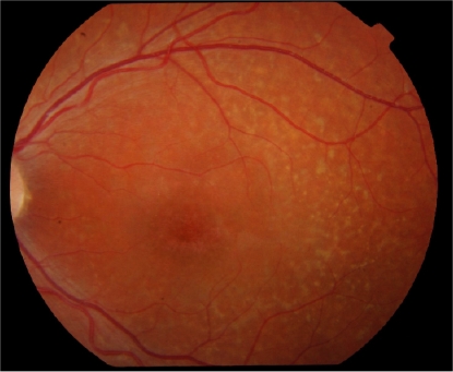
Degenerative diseases of the retina are among the most common causes of untreatable blindness in the world, and as many as ten million people in the United States have photoreceptor degenerative disease. While most of these patients have Age-Related Macular Degeneration (AMD), a smaller number have Stargardt’s, an Orphan disease and to date an untreatable form of juvenile macular degeneration leading to blindness in a much younger group of patients than are affected by AMD. ACT’s treatment for eye disease uses stem cells to re-create a type of cell in the retina that supports the photoreceptors needed for vision. These cells, called retinal pigment epithelium (RPE), are often the first to die off in SMD and AMD, which in turn leads to loss of vision.

While there is currently no treatment for SMD, several years ago ACT and its collaborators discovered that human embryonic stem cells could be a source of RPE cells. Subsequent studies found that the cells could restore vision in animal models of macular degeneration. In a Royal College of Surgeons (RCS) rat model, implantation of RPE cells resulted in 100% improvement in visual performance over untreated controls, without any adverse effects. The cells survived for more than 220 days and sustained extensive photoreceptor rescue. Functional rescue was also achieved in the ‘Stargardt’s’ mouse with near-normal functional measurements recorded at more than 70 days.
Enhanced by Zemanta

Monday, August 23, 2010

Modern Eye Prosthetics Look Real!

An ocular prosthesisImage via WikipediaOnce, artificial eyes looked patently false but the latest prosthetics used today look almost real, with precise colour-matching, accurate shaping and some eye movement possible.

Dr M. Ashwin Reddy, consultant paediatric ophthalmologist at Barts and the London and Moorfields Eye Hospital, says: 'Artificial eyes have been used for more than 100 years, first appearing in the late 19th Century'.

In the past, patients simply had a shell without anything behind to increase the volume in the eye socket. This resulted in drooping of the eyelid and didn't really make for a very good cosmetic result.
Now a patient will usually have a ball-shaped orbital implant surgically inserted into the socket. If you remove the eye, there's no tissue left, so the implant helps keep the structure of the eye socket, replaces volume and gives a better result.
This is particularly important with young children, as the bones of the eye socket can then grow around the implant.

Nigel Sapp, chief ocularist (prosthetic expert) at Moorfields Eye Hospital, explains, today's prostheses are very realistic.
'Prostheses are moulded to a patient's socket or eye remnant and the colour of the iris is slowly built up, literally using a paintbrush and some dry-powder pigment paints in front of the patient until we achieve a good colour match.
'Before, artificial eyes would come from stock. Even the white of the eye is colour-matched and vessels and veins are added using embroidery threads, to match the patient's own characteristics.'


Enhanced by Zemanta

Thursday, August 19, 2010

Radioactive discs in the eyes

Retinoblastoma retina scan before and after ch...Image via WikipediaJ. William Harbour, MD at Washington University School of Medicine in St. Louis is implanting radioactive discs in the eye as a treatment option for a rare type of cancer called retinoblastoma.

Retinoblastoma, as the name suggests, is characterized by tumors in the eye's retina. It is rare, affecting about one child in 20,000. In the United States, about 200 children each year are diagnosed with it. Approximately 40 percent of them develop tumors in both eyes, so in cases where the tumors prove resistant to chemotherapy, very young children and their parents are faced with a choice between a life without eyes and a high risk of death.

"The treatment plaque looks like a bottle cap made of gold," Harbour says. "Radiation seeds are placed on one side of the plaque, shining the radiation in one direction like a flashlight focused on the tumor. That prevents the radiation from affecting other parts of the body."

"The radiation causes damage within the cancer cells that prevents them from proliferating and spreading," he says. "By the time we take off the plaque, the cancer cells are either dead or mortally wounded, even though we do not immediately see a difference in the appearance of the tumor. After the plaque therapy, as the cancer cells try to proliferate and divide, those cells die, which we then notice in follow-up exams as the tumor shrinks over time."
Enhanced by Zemanta

Thursday, August 12, 2010

Proliferative retinopathy Description: Prolife...Image via WikipediaThe Los Angeles Times reports, Nearly three in 10 Americans with diabetes over the age of 40 suffer from vision impairment caused by diabetic retinopathy, and about 4.4% have a form of the condition so severe it threatens their eyesight, according to a study published in the August 11 issue of the Journal of the American Medical Association.

HealthDay reported that the news wasn't all bad, however. The findings suggest that good control of blood sugar, blood pressure, and cholesterol can go a long way toward preventing or slowing diabetic eye disease. For the study, CDC researchers reviewed data from the National Health and Nutrition Examination Survey from 2005 to 2008, which included nearly 7,000 people over age 40, 1,006 of whom had diabetes.

Focusing on specific gender and ethnic/racial demographics, reported that 31.6% men and 25.7% of women over age 40 who have diabetes are estimated to have diabetic retinopathy, while 38.8% of non-Hispanic blacks were estimated to have diabetic retinopathy, 34% of Mexican-Americans, and 26.4% of non-Hispanic whites. Interestingly, age was not a major factor among the study group.

Independent risk factors for diabetic retinopathy included male sex, higher hemoglobin A1c level, longer duration of diabetes, and higher systolic blood pressure, with respective odds ratios of 2.07, 1.45, 1.06 per year of duration, and 1.03 per millimeter of mercury.
Enhanced by Zemanta

Ayala carefully prepares to put a contact lensImage via WikipediaThe journal Pediatrics, in association with the Food and Drug Administration (FDA), released the results of a 2-year study looking at emergency room visits for medical device-associated adverse events in children.

Of all the visits, about 23 percent were associated with contact lens wear. These events included abrasions and ulcers of the cornea and conjunctivitis. While these did not require hospitalization, many could have been prevented.

The most common risk factors for contact lens complications in this study were alteration of the prescribed wearing or replacement schedules and non-compliance with recommended contact lens wear and care regimens. It is well known that improper care, using solutions other than those prescribed, not replacing cases or lenses as prescribed, poor hygiene, buying lenses on the Internet without a valid prescription, and not seeing the eye doctor regularly, all lead to increased risk of complications.
Enhanced by Zemanta

Tuesday, August 10, 2010

Green laser pointer can cause eye damage

astronomy: green laser pointing Orion (animation)Image via Wikipedia

Green laser pointers are readily available in stores and on the Internet. Class 3A green laser pointers are increasingly being used by amateur astronomers to pinpoint objects in the night sky and by the construction industry and architecture educators to point out details of structures in daylight.

Dennis Robertson, M.D., Mayo Clinic ophthalmologist, conducted investigations with a green laser pointer directed to the retina of a patient’s eye - scheduled for removal because of a malignancy.
The exposure was 60 seconds to the fovea, the center of acute vision; five minutes to a site 5 degrees below the fovea; and 15 minutes to a site 5 degrees above the fovea. Color photographs were taken of the eye before and after exposure to the laser.

The green laser damaged the pigment layer of the retina, although it did not cause a measurable decrease in the visual function of the patient’s eye. it is believed that longer exposures could harm vision.
Enhanced by Zemanta

Wednesday, August 04, 2010

BMI and Glaucoma

MedPage Today reported, Women with higher body mass index and weight were at reduced risk of developing certain forms of glaucoma, according to a study published in the August issue of Ophthalmology.

In fact, for each unit of body mass index, participants in the giant Nurses' Health Study showed a 6% reduction (95% CI 2% to 9%) in the rate of developing primary open-angle glaucoma with an intraocular pressure of 21 mm Hg or less at diagnosis (so-called normal tension). The study authors theorized that some measure linked to adiposity or lean mass that is under sex hormonal influences may protect against the development of primary open-angle glaucoma.

According to the researchers' report online in Ophthalmology, no relationship between body composition measures and glaucoma risk was found for men in another large cohort, the Health Professionals Follow-Up Study. Nor did BMI or other anthropometric parameters in Nurses' Health Study participants affect the risk of primary open-angle glaucoma with higher intraocular pressure.
Enhanced by Zemanta

Monday, August 02, 2010

Artificial Retina with Micro Fabrication


An implantable artificial retina fabricated at Lawrence Livermore National Laboratory is bringing sight to the blind.

“We’re talking about our progress and where we see the project going,” said Sat Pannu, who leads Livermore’s Center for Micro- and Nano-Technology, which has developed the microfabrication technology.

The project has many partners — including Second Sight Medical Products Inc., the Southern California company that has licensed the technology. Early, lower-resolution versions of the device allow people blinded for decades by the genetic eye disease Retinitis Pigmentosa or by age-related macular degeneration to see images, albeit fuzzy.

This is how it works: A miniature camera, which sits inside a pair of sunglasses, sends images wirelessly to a microprocessor worn on a patient’s belt. There it is converted to an electronic signal that is sent to a receiver on the eye, which sends the signal via a thin cable to a nanofabricated, watch-battery-size microelectrode array that is surgically tacked onto the retina. The array then emits pulses to the optic nerve, which sends a signal to the brain for processing.
The camera allows patients to zoom in and adjust the contrast of an image, Pannu said.

The project is led by Mark Humayun, a surgeon with a doctorate in biomedical engineering, at the Doheny Eye Institute at the University of Southern California. The lead laboratory is Oak Ridge National Lab in Tennessee.

Pannu, who has worked on the project since 2003, is turning again to the Department of Energy as well as the National Institutes of Health for the cash it would take to continue development through the highest-resolution version.


Enhanced by Zemanta

August is Cataract Awareness Month

Cataract Falls Upper FallsImage by W9NED via Flickr

1. A large or high waterfall.
2. A great downpour; a deluge.
3. Pathology Opacity of the lens or capsule of the eye, causing impairment of vision or blindness.


August is Cataract Awareness Month – a month when eye care professionals hope to spread the word of the causes of cataracts, the prevention and the treatments.
Cataracts are often considered an older person’s problem. It’s estimated that half of all people 80 years or older have at least one cataract. Over 20 million Americans over the age of 40 have cataracts. Although they are more common in the elderly, they’re not rare in younger people either.

The National Eye Institute identifies the different types of cataracts:

  1. Secondary cataract. Cataracts can form after surgery for other eye problems, such as glaucoma. Cataracts also can develop in people who have other health problems, such as diabetes. Cataracts are sometimes linked to steroid use.
  2. Traumatic cataract. Cataracts can develop after an eye injury, sometimes years later.
  3. Congenital cataract. Some babies are born with cataracts or develop them in childhood, often in both eyes. These cataracts may be so small that they do not affect vision. If they do, the lenses may need to be removed.
  4. Radiation cataract. Cataracts can develop after exposure to some types of radiation.
Cataracts can often be detected in the early stages and this is one of the reasons why regular eye exams are important. There are ways to help improve vision with lighting, magnifying tools and even anti-glare sunglasses until surgical removal of the cataract is warranted.

Help yourself

Although it might not be possible to avoid cataracts altogether, it is possible to limit your risk of developing one or to hold off development. To reduce your risk:

  • Don’t smoke
  • Avoid exposing your eyes to direct sunlight (use good sunglasses)
  • Drink alcohol moderately
  • Eat a diet rich with fruit and vegetables


Enhanced by Zemanta

Sunday, August 01, 2010

Tribal Clinic Working On Telemedecine

Low-resolution image of the Oregoh Health and ...Image via Wikipedia

Yellowhawk Tribal Health Center near Pendleton, OR, is one of two tribal clinics (the other is in Wichita, Kan.) working on a test of long-distance eye care with researchers from Oregon Health & Science University and Legacy Health's Devers Eye Institute. This effort waqs highlighted by The Oregonian

At Yellowhawk, patients with diabetes can undergo a retinal scan with a $20,000-plus non-mydriatic camera, which can photograph the retina without dilating the pupil. Next, the images are uploaded to the Internet and sent to doctors in Portland who check for signs of retinopathy. The images also allow doctors to check for glaucoma or macular degeneration.
Enhanced by Zemanta

Twitter Delicious Facebook Digg Stumbleupon Favorites More

 
Design by Free WordPress Themes | Bloggerized by Lasantha - Premium Blogger Themes | Bluehost Review